"Behavioral Disease Ecology Across Scales from Individuals to Populations"
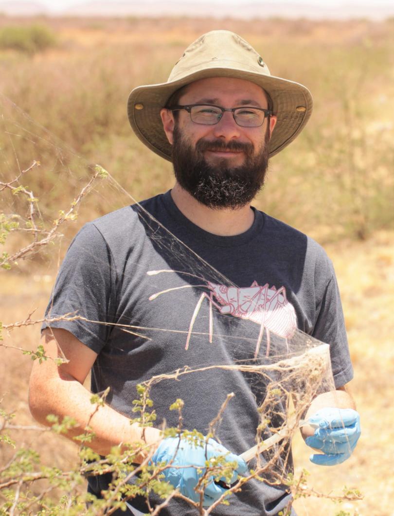 Dr. Nick Keiser | Keiser Lab
Bio:
Nick Keiser is a behavioral disease ecologist interested in how behavioral trait variation can influence infectious disease dynamics. His mentees study questions at the nexus of animal behavior, parasitology and disease ecology in a variety of such (mostly invertebrate) study systems as flies, spiders, ticks, snails and their associated parasites.
Abstract:
The fields of animal behavior and infectious disease are both typified by heterogeneity. Differences among individuals, between social groups and between populations in their behavioral trait compositions can all alter the dynamics of infectious diseases. In this seminar, I will address how animal behavior can alter host-pathogen interactions across different scales from individuals to populations in several study systems. These study systems will weave three tales on behavioral trait diversity, behavioral parasitology, and parasite manipulation of host behavior.
Watch the seminar here!